5 facts about … animal physiotherapy
Dr Naomi Boyd, Emma Mathlin, Kristin Dean, Lynne Harrison and Sally Elsom contribute five discussion points about the presentation of common conditions in equine and canine athletes and companions.
1. Cruciate rupture is common in canine companions
The cranial cruciate ligament (CCL) is the anatomical equivalent of the anterior cruciate ligament, and partial or complete tears are the most common stifle (canine knee) injury in dogs (Cook 2010). For those that rupture a CCL, 50 per cent will go on to tear their contralateral ligament within two years (Muir et al 2010). The pathophysiology of canine CCL tears are complex, the result of an interplay of elements including genetic, biomechanical, environmental and immune-mediated factors (Vasseur 2003).
Treatment also varies, and ligament replacement surgery is rarely effective in dogs (Muir et al 2010, Aragon & Budsberg 2005). Instead, veterinary cruciate surgeries attempt to change the biomechanics of the stifle, making the restraint of cranial tibial translation unnecessary (Vasseur 2003).
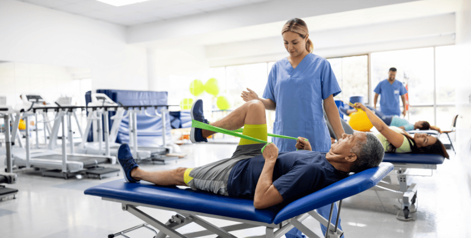
As in humans, cruciate disease is accompanied by pain, effusion, muscle atrophy, the inability to participate in play or sport and future osteoarthritis. The disease is usually chronic in dogs and these sequelae are often more pronounced. Consequently, physiotherapy is just as important, if not more so, in returning a dog to normal activity and has been demonstrated to improve outcomes (Monk et al 2006). Rehabilitation revolves around restoring quadriceps muscle strength, developing motor control as well as reducing pain and swelling.
Hydrotherapy in an underwater treadmill is particularly useful in restoring normal gait in the early stages of recovery.
References
Cook JL. Cranial cruciate ligament disease in dogs: biology ver- sus biomechanics. Vet Surg. 2010;39:270-277.
Muir et al. Contralateral Cruciate Survival in Dogs with Unilateral Non-Contact Cranial Cruciate Ligament Rupture. Plos one 2010; 6:10.
Aragon, C. L. & Budsberg, S. C. (2005) Applications of evidence-based medicine: cranial cruciate ligament injury repair in the dog. Veterinary Surgery 34, 93-98
Vasseur PB: Stifle joint, in Slatter D (ed): Textbook of Small Animal Surgery (ed 3). Philadelphia, PA, Saunders, 2003, pp 2095–2116
Monk et al. Effects of early intensive postoperative physiotherapy on limb function after tibial plateau leveling osteotomy in dogs with deficiency of the cranial cruciate ligament. AVJR: 2006; 67:3
2. Striding ahead with motion capture
Biomechanical analysis is widely used in human athletes to evaluate and optimise performance and we are now able to apply this technology to objectively measure how the horse moves and performs. Historically, agreement between vets and other clinicians around the identification of mild lameness is poor, so the development of more objective and reliable methods of examining gait and lameness is needed (Keegan et al 2009).
High-speed video cameras with specialised software analysis allows for the measurement of variables such as the length and frequency of a horse’s stride, joint angles and speeds, and how the horse sequences each movement (Hobbs et al 2010). Inertial sensor technology produces highly accurate and precise results about the symmetry of movement between left and right limbs, as well as measuring three-dimensional back movement, allowing us to detect subtle differences within the gait or movement that the human eye can’t see (Warner et al 2010).
These technologies can be used to assess a horse’s gait to pick up early, subtle or multi-limb lameness; to monitor response to treatment, training or rehabilitation; to record a baseline of a horse’s normal gait for future comparison and to provide recommendations for improving performance. It also has the advantage of being used to see the immediate effects of interventions such as changing hoof balance, physiotherapy treatment or riding instruction.
References
Keegan et al 2009. Repeatability of subjective evaluation of lameness in horses. Equine Veterinary Journal 41(1):1-6.
Hobbs, S.J. et al (2010). Motion analysis and its use in equine practice and research. Vet. Med. Austria 97: 55-64.
Warner, S.M. et al (2010). Inertial sensors for assessment of back movement in horses during locomotion over ground. Equine Veterinary Journal 42: 417-424.
3. Sacroiliac dysfunction in the equine athlete
Poor performance and hind limb gait abnormalities are frequently seen in the horse and often attributed to sacroiliac pain, resulting from reduced neuromotor control (Goff 2014, Stubbs et al 2011). Reduced motor control is believed to cause hypo/hypermobility of the sacroiliac joint (SIJ), leading to pathological changes at the joint and asymmetries of associated muscles and/or bone (Goff et al 2008).
The role of the SIJ is to transfer forces from the horse’s hind limb to the trunk. Force transfer is believed to occur via the interconnections of ligaments and fascia (Goff et al 2008). Signs and symptoms of sacroiliac dysfunction (SID) include reduced hind limb impulsion, a plaiting hind limb gait, inability to weight bear on the affected side and asymmetry of the pelvis.
Behavioural problems may also arise, especially when ridden. Functional diagnosis relies on a combination of manual tests to assess the direction and range of SIJ motion, palpation and pain provocation tests. Management of SID in horses is based on clinical presentation and may include rest, analgesia, functional stability exercises and exercises to strengthen the back and hindquarters. Treatment of the equine SIJ requires an understanding of biomechanics and neuromotor control of the region, similar to the approach given to the human SIJ.
References
Goff, L. (2014) Manual therapy and exercise for athletic horses, in Hinchcliffe, K., Kaneps, A. & Geor, R. (ed) Equine Sports Medicine and Surgery, 2nd Edition, Edinburgh, Elsevier & Saunders, pp 1217-1224
Goff, L.M., Jeffcott, L.B., Jasiewicz, J., and McGowan, C.M. (2008) Structural and biomechanical aspects of equine sacroiliac function and their relationship to clinical disease,The Veterinary Journal, p281-293
Stubbs, N., Kaiser, H. & Clayton, H (2011) Dynamic mobilization exercises increase cross sectional area of musculus multifidus, Equine Veterinary Journal, 43, (5), pp522-529
4. A harmonious connection
 In any equestrian discipline—whether that be dressage, show jumping, flat racing or barrel racing—treating only the horse means only half of the combination is attended. Riders are looking for ‘harmony’ with their horse by being able to absorb the horse’s movement, known as an independent seat (Mackechnie-Guire et al 2018). This is achieved when the rider has good dynamic stability and flexible low back and hips.
Many riders struggle to achieve an independent seat, resulting in performance inadequacies along with pain and restriction experienced by the horse due to abnormal loading (Hampson & Randle 2015, Greve & Dyson 2012).
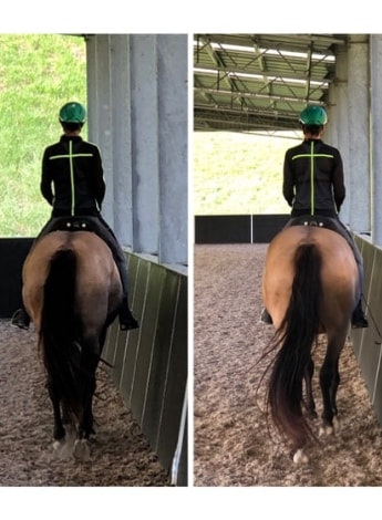
A decrease in rider gluteal and piriformis strength and overactive adductors and psoas are very common findings. Weak, spinal stabilisers will give the ‘dropped shoulder’ appearance, all leading to instability and ineffective use of the legs (Hampson & Randle 2015). In the picture left, the imbalance of the rider resulted in her being ineffective with her leg aids and the horse not being straight. Following rider treatment to release psoas and obliques, straightness and balance in the combination is achieved, along with effective leg aids.
By working with a physiotherapist, horse/rider restrictions and imbalances limiting performance can be identified and with the appropriate exercise prescription and training drills, the short comings can be overcome.
References
Mackechnie-Guire, R., Mackechnie-Guire, E., Bush R., Lawson, A., Hargreaves, S., Assirelli, S., Fairfax, V., Fisher, D., Fisher, M., and Pfau, T., (2018) Induced rider asymmetry and its effects on equine limb kinematics and thoracolumbar range of motion, ICEEP 2018, https://www.wageningenacademic.com/doi/pdf/10.3920/cep2018.s1
Greve, L., Dyson S. (2012) The horse-saddle-rider interaction, The Veterinary Journal https://www.researchgate.net/publication/257491420_The_horse-saddle-ride...
Hampson, A. & Randle, H. (2015) The Influence of an 8-week rider core fitness program on the equine back at sitting trot, International Journal of Performance Analysis in Sport, Volume 15, Issue 3, pp 1145-1159
5. Intervertebral disc disease a common cause for neurological dysfunction in dogs
Cervical intervertebral disc disease (IVDD) is characterised by low head carriage, hyperpathia to palpation, lameness and nerve root signature with or without neurological symptoms. Beagles are over-represented in the incidence of cervical IVDD. Surgical treatment involves disc decompression via a ventral slot procedure (Brisson 2010).
Thoracolumbar IVDD is the most common form of IVDD. Symptoms may include pain to spinal palpation, neurological deficits ranging from increased hind limb muscle tone or spasticity, ataxia of the hind limbs, to complete paralysis with loss of deep pain perception and an inability to urinate. Surgical management includes laminectomy and hemilaminectomy. Dachshunds are over-represented in thoracolumbar IVDD but Labradors and German shepherds have a high incidence of TL IVDD (Brisson 2010).
Lumbar and lumbosacral disc disease can present with pain and hind limb weakness (Brisson 2010). Clinical presentation is affected by the location and severity of the spinal lesion. Physiotherapy management focuses on addressing the presenting symptoms. Modalities utilised include massage and manual therapy to address pain and vertebral movement dysfunction, neuromuscular electrical stimulation, sensory and reflex stimulation, and targeted therapeutic exercise (Drum 2010). Laser therapy, when instituted early, has also been demonstrated to reduce the time to ambulation after surgery (Draper et al 2012).
References
Brisson, B. (2010). Intervertebral Disc Disease in Dogs. Veterinary Clinics Of North America: Small Animal Practice, 40(5), 829-858. doi: 10.1016/j.cvsm.2010.06.001
Draper, W., Schubert, T., Clemmons, R., & Miles, S. (2012). Low-level laser therapy reduces time to ambulation in dogs after hemilaminectomy: a preliminary study. Journal Of Small Animal Practice, 53(8), 465-469. doi: 10.1111/j.1748-5827.2012.01242.x
Drum, M. (2010). Physical Rehabilitation of the Canine Neurologic Patient. Veterinary Clinics Of North America: Small Animal Practice, 40(1), 181-193. doi: 10.1016/j.cvsm.2009.09.009
Dr Naomi Boyd, APAM, works as a sports medicine veterinarian at the Small Animal Specialist Hospital in Sydney.
APA members Kristin Dean and Emma Mathlin are the founders of Equimotion, a Sydney company specialising in physiotherapy and biomechanics of horse and rider, with a focus on using technology for objective gait and movement analysis.
Lynne Harrison, APAM, is a UK-trained physiotherapist, qualifying in 2002. She spent 13 years working in musculoskeletal physiotherapy before gaining her Master of Science in Veterinary Physiotherapy in 2015.
Sally Elsom, APAM, has been a senior physiotherapist in tertiary hospitals in the UK and Australia, working with medical and surgical specialties including critical care. She has a postgraduate diploma in veterinary physiotherapy.
Annette Bowen also provided editing and reference support for this article.
© Copyright 2024 by Australian Physiotherapy Association. All rights reserved.