
5 facts about … emergency department physiotherapy
Leigh Marchetto, Mark Cruickshank, Caitlin Farmer and Deborah Lenaghan contribute five discussion points about common presentations and occurrences in hospital emergency departments.
1. Wrist splints for buckle fractures
Torus or buckle fractures are very common paediatric injuries characterised by incomplete fracturing of the long bone after axial load being applied (Hang et al 2017). Despite these fractures being very stable and greater access to imaging and healthcare, more of these injuries are being seen at emergency departments (EDs) and outpatient care centres for definitive care/management.
The term ‘torus’ is derived from the Latin word ‘tori’ which means protuberance. A torus is the convex portion of the upper part of the base of a Greek column and resembles the appearance of the cortical buckling seen in the column of bone which has been fractured (Manaster et al 2013).
Recent guidelines and research challenge the idea that not every forearm fracture needs a cast. NICE guidelines in 2015 and systematic reviews indicate that to reduce unnecessary follow-up appointments and stiffness and pain of undisplaced (10 degrees) distal radial torus fractures, simple systems can be put into place regarding easier management (Costa 2015, Jiang 2016, Pountos et al 2010). These systems are correct identification of torus fractures, good education of parents and GPs, and the use of removable splints for 3–4 weeks (Koelink et al 2016). Not only is it helpful for hygiene for parents but it encourages more protected use of the limb, and no further imaging/medical care or excessive waits at already busy fracture clinics.
More and more EDs are using this pathway to aid patient care and reduce the burden of care, including most specialist paediatric hospitals such as the children’s hospitals at Randwick and Westmead in Sydney, and The Royal Children’s Hospital Melbourne.
References
1) B T Hang, C Gross, H Otero & R Katz (2017) An Update on Common Orthopedic Injuries and Fractures in Children: Is Cast Immobilization Always Necessary? Clinical Pediatric Emergency Medicine; 18 (1) pp 62-73
2) B. J. Manaster, D. A. May, D. G. Disler. (2013) 4th ed: Musculoskeletal Imaging, The Requisites (Expert Consult - Online and Print- Saunders),
3) M.Costa et al (2015) Fractures (non-complex): assessment and management Fractures: diagnosis, management and follow-up of fractures; National Institute for Health and Care Excellence: https://www.nice.org.uk/guidance/ng38 (accessed 3/3/19)
4) N Jiang et al (2016) Management of Pediatric Forearm Torus Fractures: A Systematic Review and Meta-Analysis Pediatric Emergency Care 32 (11) pp 773-778
5) I Pountos, J Clegg, ASiddiqui (2010) Diagnosis and treatment of greenstick and torus fractures of the distal radius in children: a prospective randomised single blind study: J Child Orthop (4):321–326
6) E. Koelink, et al (2016) Primary Care Physician Follow-up of Distal Radius Buckle Fractures PEDIATRICS Volume 137, number 1, pp 1-9

2. Bedside predictors and tests can help identify vertigo
It’s no secret that vertigo is one of the most common presentations to EDs, and one of the most complex diagnoses. With the cohort of patients that present to EDs, staff (eg, doctors, physiotherapists) ‘must differentiate vertigo caused by central nervous system pathology … from that caused by disorders of the peripheral’ vestibulopathy (Quimby et al 2018). Accessing imaging and pathology takes time and limitations on accuracy, hence ‘using bedside predictors are essential to identify patients with acute central vestibulopathies’ (Quimby et al 2018, Kattah et al 2009).
Tests such as HiNTs examination are more sensitive and specific for differentiating between central and peripheral causes in the first 48 hours of symptoms than an MRI or CT. After this, central adaptive processes take over and the HiNTs is no longer as sensitive (Kattah et al 2009). The HiNTs should only be completed in those who have continuous vertigo (eg, for hours or days). Importantly it has been demonstrated that the HiNTS exam is under-utilised in the ED as compared to neuroimaging in the assessment of patients with peripheral vertigo (Quimby et al 2018).
While peripheral pathologies such as benign positional paroxysmal vertigo (BPPV) and neuritis are common acute vestibulopathy (Tarnutzer et al 2011) in the ED, using a simple battery of tests (eg, head impulse test, nystagmus and test of skew) can help with differentiation, allow for clearer diagnosis and help with education provided on the return-to-normal activities (Quimby et al 2018, Sealy 2014). With the understanding of gait and assessment, physiotherapists are well placed to aid in diagnosis or ‘could help reduce frontline misdiagnosis of patients with stroke’ (Kattah et al 2009).
References
Quimby et al (2018) Usage of the HINTS exam and neuroimaging in the assessment of peripheral vertigo in the emergency department: Journal of otolaryngology - head & neck surgery; 47(1)
Kattah et al. (2009) HINTS to Diagnose Stroke in the Acute Vestibular Syndrome: Three-Step Bedside Oculomotor Examination More Sensitive Than Early MRI Diffusion-Weighted Imaging; Stroke; 40 pp 3504-3510.
Tarnutzer et al. (2011) Does my dizzy patient have a stroke? A systematic review of bedside diagnosis in acute vestibular syndrome: CMAJ, 183(9)
Sealy, (2014)
3. There are rules to determine the need for radiographs in acute knee and ankle injuries
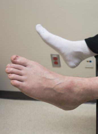
The Ottawa ankle and knee clinical prediction rules were designed to reduce unnecessary knee and ankle imaging in an acute setting. The Ottawa ankle rules were first developed by Stiell et al (1992) and are listed below.
An ankle series is required if pain is near the malleoli together with one or more of these findings: unable to weight bear both immediately and in the ED (four steps) and bone tenderness at the posterior edge or tip of either malleoli. A foot series is required if there is pain in the midfoot together with one or more of these findings: unable to weight bear both immediately and in the ED (four steps), boney tenderness of the navicular, boney tenderness of the base of the fifth metatarsal.
The Ottawa knee rules were developed by Stiell et al (1993) and are listed below. A knee series is required for acute knee injuries together with one or more of these findings: aged 55 years or older, tenderness at head of fibula, isolated tenderness of patella, inability to flex to 90 degrees, inability to bear weight both immediately and in the ED (four steps).
The Ottawa clinical prediction rules have been validated in the adult population for both the knee (Bachmann et al 2004) and ankle (Bachmann et al 2003). The Ottawa clinical prediction rules have also been validated for use with children aged over five for both the knee (Vijayasankar et al 2009) and ankle (Dowling et al 2009).
4. Low back pain is a common presentation to EDs
Low back pain is one of the most common presentations to EDs in Australia and worldwide, accounting for over four per cent of all ED complaints (Edwards et al 2017). People present to EDs with low back pain for a variety of reasons, including limited access to primary care and levels of distress and disability, and they often have high expectations of analgesia and imaging (Strudwick et al 2018).
Although only around 6.7 per cent of these patients have a serious or specific condition (Thiruganasambandamoorthy et al 2014), around a third of patients presenting to ED get some type of imaging (Downie et al 2019).
Imaging is associated with increased healthcare costs and utilisation following an episode of low back pain, and does not appear to aid recovery (de Schepper et al 2016, Graves et al 2012a, Graves et al 2012b, Lemmers et al 2019). Cross-sectional imaging, such as CT and MRI scans, are likely to show structural changes that are not necessarily the source of symptoms. For example, disc protrusions are present in a third of 40 year olds without pain (Brinjikji et al 2015). Once serious and specific conditions have been excluded, imaging is not recommended in acute low back pain and the focus should be on pain control and restoration of function (Strudwick et al 2018), basic elements of physiotherapy practice.
References
Edwards J, Hayden J, Asbridge M, Gregoire B, Magee K. Prevalence of low back pain in emergency settings: a systematic review and meta-analysis. BMC Musculoskelet Disord. 2017;18(1):143.
Strudwick K, McPhee M, Bell A, Martin-Khan M, Russell T. Review article: Best practice management of low back pain in the emergency department (part 1 of the musculoskeletal injuries rapid review series). Emerg Med Australas. 2018;30(1):18-35.
Thiruganasambandamoorthy V, Turko E, Ansell D, Vaidyanathan A, Wells GA, Stiell IG. Risk factors for serious underlying pathology in adult emergency department nontraumatic low back pain patients. J Emerg Med. 2014;47(1):1-11.
Downie A, Hancock M, Jenkins H, Buchbinder R, Harris I, Underwood M, et al. How common is imaging for low back pain in primary and emergency care? Systematic review and meta-analysis of over 4 million imaging requests across 21 years. Br J Sports Med. 2019.
de Schepper EI, Koes BW, Oei EH, Bierma-Zeinstra SM, Luijsterburg PA. The added prognostic value of MRI findings for recovery in patients with low back pain in primary care: a 1-year follow-up cohort study. Eur Spine J. 2016;25(4):1234-41.
Graves JM, Fulton-Kehoe D, Martin DP, Jarvik JG, Franklin GM. Factors associated with early magnetic resonance imaging utilization for acute occupational low back pain: a population-based study from Washington State workers' compensation. Spine (Phila Pa 1976). 2012;37(19):1708-18.
Graves JM, Fulton-Kehoe D, Jarvik JG, Franklin GM. Health care utilization and costs associated with adherence to clinical practice guidelines for early magnetic resonance imaging among workers with acute occupational low back pain. Health Serv Res. 2014;49(2):645-65.
Lemmers GPG, van Lankveld W, Westert GP, van der Wees PJ, Staal JB. Imaging versus no imaging for low back pain: a systematic review, measuring costs, healthcare utilization and absence from work. Eur Spine J. 2019.
Brinjikji W, Luetmer PH, Comstock B, Bresnahan BW, Chen LE, Deyo RA, et al. Systematic literature review of imaging features of spinal degeneration in asymptomatic populations. AJNR Am J Neuroradiol. 2015;36(4):811-6.
5. Opioid poisoning hospitalisations are on the rise
Internationally, the rising use of opioids is a cause of concern. All opioids, including codeine, can be addictive, and their use can result in dependence, accidental overdose, hospitalisation or death.
The Australian Institute of Health and Welfare (2018) found that in Australia in 2016–17, 3.1 million people had one or more prescriptions dispensed for opioids, about 40,000 people used heroin, and about 715,000 people used painkillers/analgesics and pharmaceutical opioids for illicit or non-medical purposes.
The Institute also found that opioid deaths and poisoning hospitalisations have increased in the last 10 years. Every day in Australia, nearly 150 hospitalisations and 14 ED presentations involve opioid harm—three people die from drug-induced deaths involving opioid use. In 2016, opioid deaths accounted for 62 per cent of all drug-induced deaths.
The Institute says pharmaceutical opioids are responsible for more opioid deaths and poisoning hospitalisations than heroin. In 2016, the most commonly mentioned in opioid deaths was naturally derived opioids (eg, oxycodone, codeine and morphine), which was mentioned in 49 per cent of opioid deaths. The rate of hospitalisation due to naturally derived opioids was more than twice as high as for those by heroin.
Oxycodone and codeine are the most commonly dispensed opioids, according to the Institute’s publication Opioid harm in Australia: and comparisons between Australia and Canada (tinyurl.com/yydxm6d2). The publication states that higher rates of oral morphine equivalents for opioids are dispensed in regional areas compared to very remote areas, and that one in 10 Australians have used a type of opioid for illicit or non-medical purposes, with most using pharmaceutical opioids rather than illegal opioids. Almost 10 per cent (9.7 per cent) have used painkillers/analgesics compared with 1.3 per cent who have used heroin.
Reference
AIHW (2018). Opioid harm in Australia and comparisons between Australia and Canada. A. I. o. H. a. Welfare.
Leigh Marchetto, APAM, is part of the executive committee with NSW ED network. Leigh has worked at Liverpool Hospital’s emergency department for the last seven years.
APA Musculoskeletal Physiotherapist Mark Cruickshank is a senior physiotherapist (emergency department) at Flinders Medical Centre. Mark also works privately.
APA Musculoskeletal Physiotherapist Caitlin Farmer is an Advance Practice Physiotherapist (Emergency and Neurosurgery) at St Vincent’s Hospital Melbourne.
Deborah Lenaghan, APAM, is the team leader for emergency department physiotherapists with Gold Coast Hospital and Health Service. Deborah is the chair of the APA Emergency Department group and is a current prescriber investigating the safety of physiotherapy prescriber’s research trial within Queensland Health.
© Copyright 2024 by Australian Physiotherapy Association. All rights reserved.