Diagnostic accuracy of lung ultrasound
The findings of this systematic review question the clinical role of chest X-ray as the preferred diagnostic tool for assessing lung consolidation and pleural effusion in intubated and mechanically ventilated patients in critical care settings. Q&A with Louise Hansell.
In intensive care patients, what sort of lung problems are amenable to treatment by physiotherapy interventions?
There are a plethora of lung conditions that a physiotherapist may be presented with, and with mechanically ventilated patients in intensive care, the ability to differentially diagnose between possible pathologies is vital to guide appropriate treatment delivery.
Typically, physiotherapists focus treatment on lobar collapse, which typically occurs as a result of prolonged mechanical ventilation or due to surgery, secretion retention and poor ventilation.
The treatments that we select or prescribe are directly related to our assessment findings, and so accurate assessment is important to ensure appropriate treatment selection.
Traditionally, physiotherapists might diagnose these with a stethoscope or by looking at a chest radiograph. What is wrong with those approaches?
With research bringing into question the effectiveness of chest physiotherapy treatments, it is important that we establish the accuracy of the conventional clinical tools and outcome measures used in assessment, diagnosis and review.
Our systematic review demonstrates the greater diagnostic accuracy of lung ultrasound compared with conventional tools (lung auscultation and chest radiographs) in diagnosing pathologies that are pertinent to respiratory physiotherapists in the intensive care setting in mechanically ventilated patients.
We have compared lung ultrasound against thoracic CT, which is the ‘gold standard’ when it comes to diagnosing respiratory pathology.
The limitations of our conventional respiratory assessment tools are significant.
Chest radiographs are often taken daily in an environment like intensive care, looking at worsening or improvement of lung pathologies day to day, but as we know, this comes with a financial cost, as well as daily exposure to radiation for patients.
Images will also only give us a snapshot in time, so to speak, of a patient’s lungs, and don’t take into account the dynamic nature of lung tissue.
The quality of an image is dependent on the skill of the radiographer and the interpretation of the image dependent on the skill of the clinician.
Physiotherapists have a long history of relying on lung auscultation.
A stethoscope is cheap and can go anywhere with you, giving us accessible ‘real time’ information regarding aeration of lung tissue.
Unfortunately, auscultation has poor intra and inter-rater reliability, and the interpretation of sounds is highly subjective and dependent on the skill of the clinician.
We found the sensitivities and specificities of lung auscultation for various pathologies, reported by only one study in our systematic review, were highly varied.
If we consider the low diagnostic accuracy of these tools, we acknowledge that our clinical impression and associated treatment plans are based on potentially inaccurate clinical information, and so may not be correct or entirely appropriate.
If conventional diagnostic tools have low diagnostic accuracy, and physiotherapists can incorporate assessment tools with higher accuracy, we may be better able to select appropriate treatment and improve patient outcomes.
Your systematic review compared the diagnostic accuracy of those approaches against lung ultrasound. Does lung ultrasound avoid the limitations of those other methods?
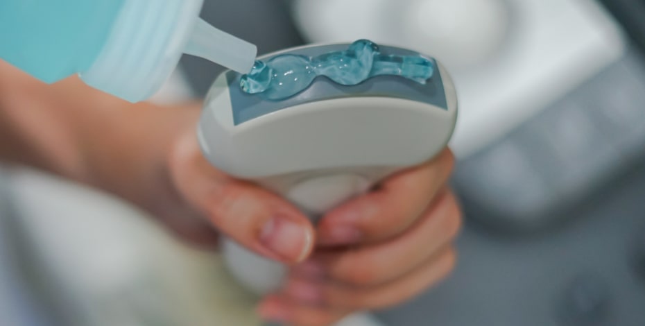
The great thing about lung ultrasound is that it ticks many boxes in relation to what we are looking for in an assessment tool.
It is portable and able to be moved from the bedside of one patient to the next easily.
Unlike chest X-ray, it is radiation-free, which makes it safe for patients to undergo multiple ultrasound investigations daily if ever required.
A basic model is quite affordable, with minimal ongoing cost, only those associated with consumables like ultrasound gel.
Most hospitals would have ultrasound units already available for use, which would eliminate the need to purchase any equipment.
There is also no pain associated with an ultrasound assessment as it is a non-invasive procedure.
With the high diagnostic accuracy of lung ultrasound that we reported in the systematic review, when compared with chest radiograph and auscultation, it has potential to improve physiotherapists’ capacity to better diagnose and treat respiratory impairments.
Performing and interpreting a lung ultrasound as a physiotherapist does require training, however once you have attained these skills, performance and interpretation of a full assessment on a single patient could take as little as 10 minutes.
Of note, lung ultrasound cannot diagnose airway issues, for example secretion retention or bronchospasm (a stethoscope is required for that), hence ultrasound should be part of a clinicians tool box.
How much evidence did you find?
There was a small number of studies that reported the diagnostic accuracy of lung ultrasound compared with conventional assessment tools specifically in relation to lung consolidation/collapse and pleural effusion.
This is likely a direct result of lung ultrasound being a novel and emerging tool that is not commonly used in intensive care at present.
Additionally, our inclusion criteria was very narrow, with a specific focus on these pathologies in mechanically ventilated adult patients in intensive care.
There are many other studies looking at the diagnostic accuracy of lung ultrasound for other pathologies; however, we narrowed our selection down to these specific pathologies as we felt they were the most pertinent to physiotherapists, and differentiating between these pathologies is vital to determine an appropriate treatment course.
Our systematic review found seven studies in total, five of those were able to be included in the meta-analysis, with a sample size of 253 participants.
We are hoping our study highlights lung ultrasound as a potential new assessment tool for physiotherapists.
How did lung ultrasound compare to chest radiograph?
We demonstrated that lung ultrasound had a high sensitivity (92 and 91 per cent) when compared with chest radiograph (53 and 42 per cent) when diagnosing lung consolidation/collapse and pleural effusion respectively.
The two tests had similar specificities when diagnosing both pathologies (lung ultrasound 92 per cent and chest radiograph 78 and 81 per cent respectively).
Lung ultrasound also had a high diagnostic odds ratio and area under the curve, and strongly diagnostic positive and negative likelihood ratios.
All of these diagnostic accuracy measures indicate that lung ultrasound is more discriminatory in diagnosing consolidation or effusion, and is a more suitable diagnostic tool than chest radiographs for such pathologies.
And what about auscultation with a stethoscope?
Lung auscultation was not able to form part of our meta-analysis, as only one study reported data relating to auscultation.
This study reported sensitivities of 8 and 42 per cent and specificities of 100 and 90 per cent in the diagnosis of lung consolidation and pleural effusion respectively.
This supports literature and anecdotal evidence that suggests auscultation is very subjective and dependent on the skill of the clinician carrying it out.
When we compare these sensitivities in particular with those of lung ultrasound, the numbers really do speak for themselves.
Lung ultrasound has a much greater ability to correctly identify lung consolidation and pleural effusion over lung auscultation.
Do you foresee physiotherapists using ultrasound with every respiratory treatment in intensive care units in the future?
Another great question. We are yet to determine how best to incorporate lung ultrasound into physiotherapy clinical assessment, predominately because it is still an emerging modality and not widely used at present.
There are many issues surrounding implementation of lung ultrasound into clinical practice that need further investigation, including governance, delivery of training and gaining competence and accreditation.
Additionally, consideration has to be given as to whether lung ultrasound would be considered as an extended scope or advanced scope of practice tool, or something that could form part of everyday clinical assessment for all clinicians.
The important part of our research to consider is that we are not setting out to replace conventional assessment tools, but we want to add something in to our assessment repertoire that improves our diagnostic ability as clinicians.
Anecdotally, we have seen lung ultrasound to be of the most benefit in the assessment and management of the more complex intensive care patients, new patients with complex care needs, those who have perhaps not responded to chest physiotherapy or those who continue to deteriorate from a respiratory point of view.
In saying that, any patient can undergo a lung ultrasound; its portability, affordability and safety make it the ideal assessment tool.
Additional studies that form part of my PhD will also prove valuable in highlighting the profile of lung ultrasound for physiotherapists, and demonstrate its practical applications.
I am currently undertaking an observational study at RNSH in ICU that uses lung ultrasound to detect aeration change associated with chest physiotherapy treatment in a mechanically ventilated population with atelectasis, as well as determining whether there is correlation between lung ultrasound when compared to lung auscultation, chest radiographs and clinical measures including arterial blood gas analysis and ventilator settings.
My clinical supervisor, and an expert in lung ultrasound, Dr George Ntoumenopoulos, is also collaborating with our colleague in Paris, Aymeric Le Neindre, on an exciting study in several ICUs in Paris and at St Vincent’s Hospital, Sydney investigating whether the addition of lung ultrasound to measure lung aeration into clinical assessment will alter clinical decision-making in the delivery of chest physiotherapy and clinical decisions by intensivists.
>> Louise Hansell is a physiotherapist at Royal North Shore Hospital, Sydney, and is completing a PhD through the University of Sydney focusing on lung ultrasound for physiotherapists. Louise has a particular interest in respiratory physiotherapy in the intensive care and postoperative settings.
© Copyright 2024 by Australian Physiotherapy Association. All rights reserved.