Physiotherapy and cruciate ligament disease in dogs
Katrinka Geelen, APAM, presents five discussion points about cranial cruciate ligament disease in dogs.
1. Dogs are susceptible to cruciate ligament injuries
Cranial cruciate ligament disease (CCLD) is the leading cause of hindlimb lameness in the dog (Powers et al 2005).
The cranial cruciate ligament (CCL) is the canine equivalent of the human anterior cruciate ligament.
Cranial cruciate ligament disease is usually caused by chronic degenerative change.
The CCL is the primary passive stabiliser of the canine stifle joint, its craniomedial band taut during flexion, while the caudolateral band and lateral collateral ligament relax to allow for tibial internal rotation (de Rooster et al 2006).
In addition to stifle joint stability, these ligaments provide mechanoreceptor and proprioceptive input (de Rooster et al 2006).
Injuries of the human anterior cruciate ligament typically result from acute, non-contact trauma involving deceleration or acceleration in combination with a knee valgus load (Logerstedt et al 2010).
Acute CCL rupture in dogs can result from a single high-load traumatic event; however, in most cases CCLD is the result of normal forces placed on a ligament that has undergone chronic degenerative change (Jerram & Walker 2003, Hayashi et al 2003).
This process is progressive, involving gradual degeneration of the CCL and stifle joint inflammation, and leads to partial and eventually complete ligament rupture (Hayashi et al 2003).
Secondary osteoarthritis frequently develops (de Bruin et al 2007).
2. Identifying risk factors is important in managing cruciate ligament disease
The aetiopathogenesis for CCLD is complex and not completely understood (Comerford et al 2011).
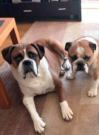
A variety of dog breeds are affected, with the highest prevalence seen in the Newfoundland, Rottweiler, Labrador Retriever, Bulldog and Boxer (Witsberger et al 2008).
A genetic basis for inheritance has been identified in Newfoundlands (Wilke et al 2006).
A number of risk factors for CCLD include:
- an increased prevalence in female dogs and in neutered over sexually intact dogs (Slauterbeck et al 2004)
- obesity, with overweight dogs demonstrating altered kinematics relative to healthy-weight dogs (Lapman et al 2003, Bray et al 2013)
- medial patella luxation, genu varum and conformational variations such as a straight stifle joint angle, narrow distal femoral intercondylar notch and steep tibial plateau angle (TPA) (Haynes et al 2015, Schwandt et al 2006, Mostafa et al 2009)
- age, given that material properties of the CCL decrease with age, increasing the risk of rupture in dogs aged more than four years, especially in ‘high risk’ breeds (Witsberger et al 2008, Duval et al 1999)
- breed variation in biomechanical properties of the CCL, with significantly lower load-to-failure properties reported in some breeds (Wingfield et al 2000a, Wingfield et al 2000b)
- immune-mediated conditions of the stifle (Doom et al 2008).
Despite the multiple risk factors for CCLD, it remains unclear which of these lead to disease initiation (Hayashi 2018).
3. Extracapsular techniques can be effective in cruciate ligament repair
Lateral fabellar stabilisation (LFS) (also known as the De Angelis procedure) is an extracapsular repair technique (De Angelis & Lau 1970).
The fabellae are small sesamoid bones, one for each tendon of the two heads of gastrocnemius (Kowaleski et al 2018).
There are a number of risk factors for cranial cruciate ligament disease, including sex, age, weight and breed.
With LFS, the aim is to passively stabilise the stifle and prevent cranial (anterior) tibial translation with respect to the femur, which would normally be prevented by the intact CCL (Heffron & Campbell 1978).
A non-absorbable synthetic suture material is passed around the lateral fabella and back through one or two bone tunnels made in the tibial tuberosity (Casale & McCarthy 2009).
The suture is held taut by a knot or crimp clamp (Lotsikas et al 2013).
The suture material eventually stretches or breaks, but the temporary stabilisation it provides allows for the formation of organised scar tissue, ultimately leading to long-term stability (Warzee et al 2001).
The advantages of LFS are its relative technical ease and shorter surgical time; however, low tensile strength or increased creep of the suture material can lead to insufficient or excessive scar tissue formation and joint laxity (Christopher et al 2013).
4. Corrective osteotomies change biomechanics but provide long-term stability
Tibial plateau levelling osteotomy (TPLO) and tibial tuberosity advancement (TTA) are the two most common corrective osteotomy techniques performed (Conzemius et al 2005).
TPLO involves cutting a curved osteotomy through the proximal tibia and rotating the tibial plateau segment to achieve a lower TPA (Lotsikas et al 2013).
After rotation, a plate and screws are applied to stabilise the osteotomised segment (Kowaleski et al 2018).
The aim is to neutralise cranial drawer by levelling the tibial surface so it is perpendicular to the load applied during weight-bearing (Kowaleski et al 2018).
TPLO alters biomechanics by placing additional reliance on the caudal cruciate ligament and muscular stabilisers of the stifle.
TTA involves an osteotomy caudal to the tibial tuberosity and advancing of the tibial tuberosity so the patellar tendon is perpendicular to the tibial plateau.
A titanium cage and plate hold the advanced portion of bone in place (Kowaleski et al 2012).
TTA neutralises cranial drawer by altering forces through the stifle to be parallel to the patellar tendon, providing a biomechanical advantage to the quadriceps muscles (Guerrero et al 2011).
This procedure is less technically demanding but has a higher incidence of postoperative patellar tendonitis and meniscal tears compared with TPLO (Hurt et al 2011).
More recent studies have shown that TPLO and TTA are superior to extracapsular stabilisation techniques, with TPLO providing better long-term radiographic and functional outcomes than TTA (Krotscheck et al 2016, Moore et al 2020).
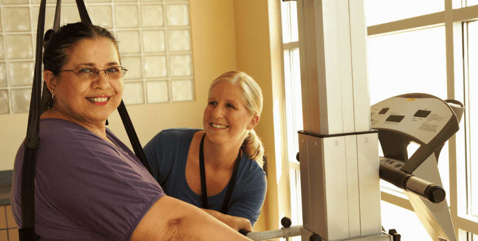
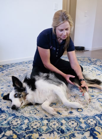
5. Physios play an important role in postoperative outcomes
Physiotherapy following CCL stabilisation has been reported in three studies (Marsolais et al 2002, Monk et al 2006, Jerre 2009).
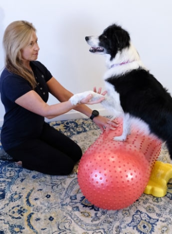
Physiotherapy is important for recovery after surgical stabilisation of the cranial cruciate ligament.
Marsolais and colleagues (2002) assessed the effectiveness of passive range of motion (PROM), massage, walking and swimming following extracapsular stifle stabilisation and found improvements in peak vertical force and vertical impulse six months after surgery.
Monk et al (2006) compared the physiotherapy modalities of cold, massage, PROM, underwater treadmill and progressive functional exercises to a home exercise program in dogs following TPLO.
At six weeks thigh circumference and stifle PROM was significantly greater in the physiotherapy group compared with controls (Monk et al 2006).
Jerre (2009) compared two groups, with the control group receiving a graduated walking program, with instructions for massage and stretching provided to the owner.
In addition, the treatment group received swimming and transcutaneous electrical nerve stimulation.
No significantly detectable differences were reported between the two groups.
It has been suggested that physiotherapy following CCL stabilisation may be more important in terms of overall patient outcome than the choice of surgical technique (Marcellin-Little & Arnoldy 2018).
Click here for an infographic poster version of this article.
>> Katrinka Geelen, APAM, works as a physiotherapist at Queensland Veterinary Specialists. She has a postgraduate diploma in veterinary physiotherapy and is currently researching canine cruciate disease as part of her master’s degree in veterinary physiotherapy.
- References
Brady, R.B., Sidiropoulos, A.N., Bennett, H.J., Rider, P.M., Marcellin-Little, D.J., and Devita, P. (2013) ‘Evaluation of gait-related variables in lean and obese dogs at a trot’, American Journal of Veterinary Research, 74(5), pp. 757 – 762.
Casale, S.A., and McCarthy, R.J. (2009) ‘Complications associated with lateral fabellotibial suture surgery for cranial cruciate ligament injury in dogs: 363 cases (1997-2005), Journal of the American Veterinary Medical Association, 234, pp. 229.
Christopher, S.A., Beetem, J., and Cook, J.L. (2013) ‘Comparison of Long‐Term Outcomes Associated With Three Surgical Techniques for Treatment of Cranial Cruciate Ligament Disease in Dogs’, Veterinary Surgery, 42, pp. 329 – 334.
Comerford, E.J., Smith, K., and Hayashi, K. (2011) ‘Update on the aetiopathogenesis of canine cranial cruciate ligament disease’, Veterinary and Comparative Orthopaedics and Traumatology, 2, pp. 91 – 98.
Conzemius, M.G., Evans, R.B., Besancon, M.F., Gordon, W.J., Horstman, C.L., Hoefle, W.D., Nieves, M.A., and Wagner, S.D. (2005) ‘Effect of surgical technique on limb function after surgery for rupture of the cranial cruciate ligament in dogs’, Journal of the American Veterinary Medical Association, 226, pp. 232 – 236.
De Angelis, M., and Lau, R.E. (1970) ‘A lateral retinacular imbrication technique for the surgical correction of anterior cruciate ligament rupture in the dog’, Journal of the Veterinary Medical Association, 157 (1), pp. 79 – 84.
de Bruin, T., de Rooster, H., Bosmans, T., Duchateau, L., van Bree, H., and Gielen, I. (2007) ‘Radiographic assessment of the progression of osteoarthrosis in the contralateral stifle joint of dogs with a ruptured cranial cruciate ligament’, The Veterinary Record, 161, pp. 745 – 750.
De Rooster, H., De Bruin, T., and Van Bree, H. (2006) 'Morphologic and Functional Features of the Canine Cruciate Ligaments', Veterinary Surgery, 35, pp. 769–780.
Doom, M., de Bruin, T., de Rooster, H., van Bree, H., and Cox, E. (2008) ‘Immunopathological mechanisms in dogs with rupture of the cranial cruciate ligament’, Veterinary Immunology and Immunopathology, 125 (1 – 2), pp. 143 – 161.
Duval, J.M., Budsberg, S.C., Flo, G.L., and Sammarco, J.L. '(1999) 'Breed, sex, and body weight as risk factors for rupture of the cranial cruciate ligament in young dogs', Journal of the American Veterinary Medical Association, 215(6), pp.811- 814.
Guerrero, T. G., Pozzi, A., Dunbar, N., Kipfer, N., Haessig, M., Beth Horodyski, M., and Montavon, P. M. (2011) ‘Effect of tibial tuberosity advancement on the contact mechanics and the alignment of the patellofemoral and femorotibial joints’, Veterinary Surgery, 40, pp. 839–848
Haynes, K.H., Biskup, J., Freeman, A., and Conzemius, M.G. (2015) ‘Effect of Tibial Plateau Angle on Cranial Cruciate Ligament Strain: An Ex Vivo Study in the Dog’, Veterinary Surgery, 44(1), pp. 46 - 49.
Heffron, L.E., and Campbell, J.R. (1978) ‘Morphology, histology and functional anatomy of the canine cranial cruciate ligament’, Veterinary Record, 102 (13), pp. 280 - 283.
Hayashi, K., Frank, J.D., Dubinsky, C., Zhengling, H., Markel, M.D., Manley, P.A., and Muir, P. (2003) ‘Histologic changes in ruptured canine cranial cruciate ligament, Veterinary Surgery, 32, pp. 269–277.
Hayashi, K. (2018) ‘Chapter 6: Histology of Cruciate Ligament Rupture’. In: Muir, P. (ed.). Advances In The Canine Cranial Cruciate Ligament. Oxford: Wiley-Blackwell, pp. 47 - 56.
Hurt, R. J., McAbee, K., and Cavanaugh, R. (2011) ‘Incidence and risk factors of postliminary meniscal injury following tibial tuberosity advancement in 116 dogs’. In: Proceedings of the 38th Annual Conference of the Veterinary Orthopaedic Society. March 5–12, Snowmass, CO, p. 44.
Jerram, R.M., and Walker, A.M. (2003) ‘Cranial cruciate ligament injury in the dog: pathophysiology, diagnosis and treatment’, New Zealand Veterinary Journal, 51(4), pp. 149-158.
Jerre S. (2009) ‘Rehabilitation after extra-articular stabilisation of cranial cruciate ligament rupture in dogs’, Veterinary Comparative Orthopaedics and Traumatology, 22, pp. 148–152.
Kim, S.E., Pozzi, A., Kowaleski, M.P., and Lewis, D.D. (2008) ‘Tibial Osteotomies for Cranial Cruciate Ligament Insufficiency in Dogs’, Veterinary Surgery, 37, pp.111–125.
Kim, S.E., Pozzi, A., Banks, S.A., Conrad, B.P., and Lewis, D.D. (2010) ‘Effect of Cranial Cruciate Ligament Deficiency, Tibial Plateau Leveling Osteotomy, and Tibial Tuberosity Advancement on Contact Mechanics and Alignment of the Stifle in Flexion’, Veterinary Surgery, 39, pp. 363 – 370.
Kowaleski, M.P., Boudrieau, R.J., Pozzi, A. (2018) ‘Stifle Joint’. In: Johnston, S.A., and Tobias, K.M. (eds.). Veterinary Surgery Small Animal. Missouri: Elsevier, pp. 2211 – 2712.
Krotscheck, U., Nelson, S.A., Todhunter, R.J., Stone, M., and Zhang, Z. (2016) ‘Long term functional outcome of tibial tuberosity advancement vs. tibial plateau leveling osteotomy and extracapsular repair in a heterogeneous population of dogs’, Veterinary Surgery, 45, pp. 261–268.
Lapman, T.J., Lund, E.M., and Lipowitz, A.J. (2003) ‘Cranial cruciate disease: Current status of diagnosis, surgery and risk for disease’, Veterinary and Comparative Orthopaedics and Traumatology, 16, pp. 122.
Lazar, T.P., Berry, C.R., Dehaan, J.J., Peck, J.N., and Correa, M. (2005) ‘Long-term radiographic comparison of tibial plateau levelling osteotomy versus extra-capsular stabilisation for cranial cruciate ligament rupture in the dog’, Veterinary Surgery, 34, pp. 133 – 141.
Logerstedt, D.S., Snyder-Mackler, L., Ritter, R.C., Axe, M.J., and Godges, J.J. (2010) ‘Knee stability and movement coordination impairments: knee ligament sprain’, Journal of Orthopaedics and Sports Physical Therapy, 40(4):A1e37.
Lotsikas, P.J., Canapp, S.O., Dyce, J., Kirkby, K., Christopher, S., and Ridge, P.A. (2013) ‘Disorders of the Pelvic Limb: Diagnosis and Treatment’. In: Zink, M.C., and Van Dyke, J.B. (eds.) Canine Sports Medicine and Rehabilitation. Iowa: Wiley-Blackwell, pp. 267 - 295.
Marcellin-Little, D.J., and Arnoldy, C.J. (2018) 'Rehabilitation for dogs with cranial cruciate ligament rupture'. In: Muir, P. Advances in the cranial cruciate ligament (2nd ed). Ames, Iowa, pp. 343-351.
Marsolais, G.S., Dvorak, G., and Conzemius, M.G. (2002) ‘Effects of postoperative rehabilitation on limb function after cranial cruciate ligament repair in dogs’, Journal of the American Veterinary Medical Association, 220, pp. 1325 – 1330.
Monk, M.L., Preston, C.A., and McGowan, C.M. (2006) ‘Effects of early intensive post-operative physiotherapy on limb function after tibial plateau leveling osteotomy in dogs with deficiency of the cranial cruciate ligament’, American Journal of Veterinary Research, 67, pp. 529 – 536.
Moore, E.V., Weeren, R., and Paek, M. (2020) ‘Extended long-term radiographic and functional comparison of tibial plateau leveling osteotomy vs tibial tuberosity advancement for cranial cruciate ligament rupture in the dog’, Veterinary Surgery, 49, pp. 146–154.
Mostafa, A.A., Griffon, D.J., Thomas, M.W., and Constable, P.D. (2009) ‘Morphometric characteristics of the pelvic limbs of Labrador Retrievers with and without cranial cruciate ligament deficiency’, American Journal of Veterinary Research, 70(4), pp. 498 - 507.
Powers, M.Y., Martinez, S.A., Lincoln, J.D., et al. (2005) ‘Prevalence of cranial cruciate ligament rupture in a population of dogs with lameness previously attributed to hip dysplasia: 369 cases (1994-2003)’, Journal of the American Veterinary Medical Association, 227(7), pp. 1109–1111.
Schwandt, C. S., Bohorquez-vanelli, A., Tepic, S., Hassig, M., Dennler, R. Vezzoni, A., and Montavon, P.M. (2006) ‘Angle between the patellar ligament and tibial plateau in dogs with partial rupture of the cranial cruciate ligament’, American Journal of Veterinary Research, 67, pp. 1855-1860.
Slauterbeck, J.R., Pankratz, K., Xu K.T., Bozeman, S.C., and Hardy, D.M. (2004) ‘Canine ovariohysterectomy and orchiectomy increases the prevalence of ACL injury’, Clinical Orthopaedics and Related Research, 429, pp. 301 – 305.
Slocum, B., and Devine, T.D. (1993) ‘Tibial plateau leveling osteotomy for repair of cranial cruciate ligament rupture in the canine’, Veterinary Clinics in North America: Small Animal Practice, 23(4), pp. 777–795.
Warzee, C.C., Dejardin, L.C.M., Arnoczky, S.P., and Perry, R.L. (2001) ‘Effect of Tibial Plateau Leveling on Cranial and Caudal Tibial Thrusts in Canine Cranial Cruciate–Deficient Stifles: An In Vitro Experimental Study’, Veterinary Surgery, 30, pp.278 – 286.
Wilke, V.L., Conzemius, M.G., Kinghorn, B.P., et al (2006) ‘Inheritance of rupture of the cranial cruciate ligament in Newfoundlands’, Journal of the American Veterinary Medical Association, 228, pp. 61 – 64.
Wingfield, C., Amis, A.A., Stead, A.C., and Law, H.T. (2000a) ‘Cranial cruciate stability in the rottweiler and racing greyhound: an in vitro study’, Journal of Small Animal Practice, 41, pp. 193 – 197.
Wingfield, C., Amis, A.A., Stead, A.C., and Law, H.T. (2000b) ‘Comparison of the biomechanical properities of rottweiler and racing greyhound cranial cruciate ligaments’, Journal of Small Animal Practice, 41, pp. 303 – 307.
Witsberger, T.H., Villamil, J.A., Schultz, L.G., Hahn, A.W., and Cook, J.L. (2008) ‘Prevalence of and risk factors for hip dysplasia and cranial cruciate ligament deficiency in dogs’, Journal of the American Veterinary Medical Association, 232, pp. 1818 – 1824.
© Copyright 2025 by Australian Physiotherapy Association. All rights reserved.