Physiotherapy and spinal cord injury
APA Neurology group members Brooke Wadsworth, Danielle Ferguson and Christine Rimmer present five facts about physiotherapy interventions for spinal cord injuries.
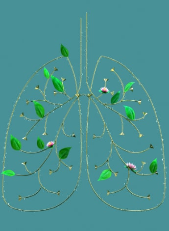
1. Airway clearance is essential for people with cervical or thoracic spinal cord injury
Respiratory complications remain the most common cause of mortality following spinal cord injury (SCI).
The ability to breathe deeply and cough forcefully is impaired to varying degrees depending on the level and completeness of the SCI.
Patients are most vulnerable to respiratory illness in the first year after injury but continue to suffer from respiratory complications throughout life (Berlowitz et al 2016).
Lung volume recruitment techniques using a modified resuscitation bag can address reduced lung capacity by augmenting the patient’s own inspiratory effort.
When used in combination with hands-on techniques (percussion or expiratory vibrations), a manual assisted cough or mechanical cough assistance device, this can create effective airway clearance (Torres-Castro et al 2014).
The ability to breathe deeply and cough forcefully can be impaired to varying degrees after spinal cord injury.
The use of a mechanical cough assistance devices that offer positive and negative pressure in addition to bilevel treatments may also be indicated to address respiratory insufficiency (Branson et al 2020).
Lung volume recruitment and cough augmentation should be taught to all self-ventilating people with a spinal cord injury who do not have fully functioning intercostal or abdominal muscles.
Failure to ensure that a patient has a preventative program to address this can increase the risk of pneumonia and respiratory failure.
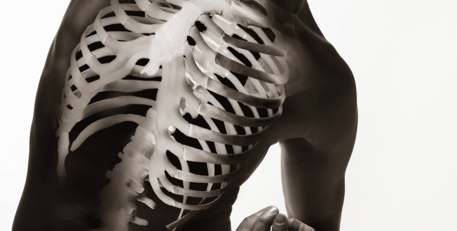
2. Musculoskeletal injury management is a key area for people with spinal cord injury
Pain is a factor for 48–94 per cent of people after SCI, with 30–40 per cent experiencing severe disabling pain that may be musculoskeletal, visceral or neuropathic (Mehta et al 2019).
Long-term wheelchair users are likely to experience upper extremity pain due to increased physical demands on these joints.
Approximately 80 per cent of individuals with chronic SCI have osteopenia or osteoporosis.
Fragility fractures occur most commonly in the distal femur or proximal tibia, susceptibility increasing with decline in bone mineral density (Svircev & Jelena 2012).
Fractures can result from a fall or with relatively no force, such as turning in bed.
There is inconclusive evidence that passive standing improves bone mineral density, although it may reduce spasticity and improve range of motion (Craven et al 2020).
Functional electrical stimulation cycling, often used for strengthening partially paralysed muscles, has other proposed benefits, such as improving general health and bone mineral density.
Heterotopic ossification occurs in 10–78 per cent of post-traumatic SCI (Alibrahim et al 2018).
This affects large synovial joints, commonly the hips.
Medications may assist with prevention and management, along with gentle stretches.
However, heterotopic ossification remains difficult to predict, prevent and treat.
3. Specific equipment prescription and management are essential for quality of life
Postural asymmetries are often associated with pain, skin breakdown and functional and respiratory impairment.
People with an SCI have an increased thoracic kyphosis, reduced lumbar lordosis and therefore posterior pelvic tilt.
Asymmetries increase the risk of a pressure injury, which 50–80 per cent of individuals with an SCI will develop in their lifetime (Hsieh et al 2020).
Long-term wheelchair use puts increased physical demands on the joints of the upper extremities.
Postural abnormalities can worsen over time if the individual is using equipment that is inappropriate or no longer meets their needs.
Intrinsic and extrinsic factors are considered when prescribing equipment.
Completing a physical examination such as the mechanical assessment tool in sitting and supine gives a better understanding of postural asymmetries and range of motion.
Photos or videos of static and dynamic postures are useful, while interface pressure mapping can be a beneficial assessment tool for examining pressure between the body and supporting surfaces.
Information on pressure variation can be used to interpret factors contributing to the development of pressure injuries.
General seating principles, as recommended by the Queensland Spinal Outreach Team, include maximising surface area contact, maintaining or improving postural control, providing a stable base of support, decreasing abnormal tone influences and promoting increased sitting tolerance (Reznik & Simmons 2020).
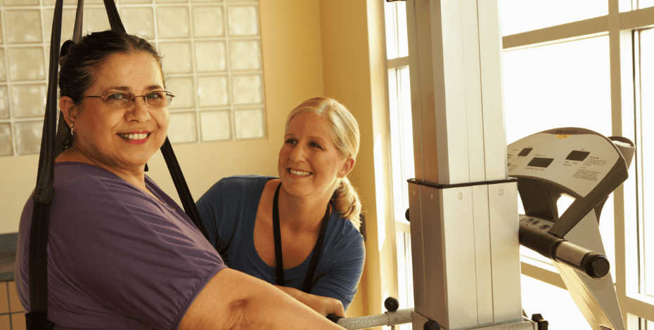
4. Autonomic dysfunction affects exercise capacity following spinal cord injury
Autonomic nervous system dysfunction can follow SCI at or above the level of T6, increasing the impact of sensory, motor and respiratory impairments on exercise capacity (Tweedy et al 2017).
Autonomic dysreflexia is the unmodulated elevation in blood pressure combined with bradycardia that occurs in response to a noxious stimulus (commonly bladder distension, constipation or painful stimulus below the level of SCI) (Middleton et al 2013).
Physiotherapists must be able to identify signs of autonomic dysreflexia (including headache, visual disturbance, light-headedness, flushed/blotchy skin and/or excessive sweating) and identify possible triggers (Middleton et al 2013).
For clients to engage safely with exercise after spinal cord injury, physiotherapists need to be familiar with the signs of autonomic dysreflexia.
Autonomic dysreflexia is an absolute contraindication to exercise and requires urgent management.
Bladder emptying prior to exercise can reduce risk, while positional changes during exercise and partaking in regular physical activity can improve tolerance (Tweedy et al 2017).
Orthostatic and exertional hypotension can also occur.
There may be a reduction in sweating, resulting in increased risk of heat stroke (Tweedy et al 2017).
Environmental controls are vital to ensuring safe engagement with exercise.
Monitoring perceived exertion is often a more reliable measure of exercise intensity than heart rate monitoring alone, especially for people with tetraplegia, as heart rate response to physical exertion is reduced (Tweedy et al 2017).
For further information related to evidence-based recommendations for physical activity after SCI, readers should refer to Physical Activity Guidelines for Adults with a Spinal Cord Injury (Martin Ginis et al 2018) and Exercise and Sports Science Australia’s position statement on exercise and SCI (Tweedy et al 2017).
5. Non-pharmacological management of spasticity following spinal cord injury
Spasticity has the potential for both positive impacts (such as reduced muscle atrophy and circulatory benefits) and negative impacts (such as interference with sleep, pain or discomfort, development of contracture and impact on functional mobility and safety).
Effective spasticity management following SCI requires a multidisciplinary approach, with evaluation of individual responses to pharmacological and non-pharmacological interventions.
The goal of management is not to eliminate the hyper-reflexive response entirely, but to overcome the impairment of function, comfort and safety (Hsieh et al 2019).
Non-pharmacological interventions leading to short-term reductions in spasticity include (Hsieh et al 2019):
- stretching—particularly when applied as a prolonged stretch
- standing—to be undertaken for 30–60 minutes at least three times per week, commencing as soon as possible following SCI (MASCIP Clinical Guideline 2019)
- splinting—via braces, thermoplastic and other positioning supports, which can also maintain and improve muscle length
- strengthening—through active exercise, both of the agonist (if voluntary motor innervation is present) and antagonist
- electrical stimulation—although there is conflicting evidence that long-term use of electrical stimulation may increase spasticity.
Overall, responses to interventions and outcomes from available evidence vary and an individualised approach that takes into account objective and subjective outcomes must be adopted for effective spasticity management.
For a comprehensive review of outcome measures specific to evaluation of spasticity following SCI, click here.
Click here for an infographic poster version of this article.
>> Brooke Wadsworth is a physiotherapist and researcher in the Queensland Spinal Cord Injuries Service at the Princess Alexandra Hospital and the Hopkins Centre, Metro South Health. Her research and clinical practice focus on rehabilitation for acute spinal cord injury and breathing dysfunction.
>> Danielle Ferguson is a physiotherapist in the Queensland Spinal Cord Injuries Service at the Princess Alexandra Hospital. Her clinical interest is rehabilitation following spinal cord injury in the inpatient and community settings. She is an active APA member for the Queensland Cardiorespiratory and Neurology special interest groups.
>> Christine Rimmer is a senior physiotherapist in the State Spinal Unit at Fiona Stanley Hospital, State Rehabilitation Service in Perth, Western Australia. Her clinical interest and specialty is inpatient rehabilitation following spinal cord injury.
- References
1. Berlowitz, D. J., Wadsworth, B., & Ross, J. (2016). Respiratory problems and management in people with spinal cord injury. Breathe, 12(4), 328-340.
2. Torres-Castro, R., Vilaró, J., Vera-Uribe, R., Monge, G., Avilés, P., & Suranyi, C. (2014). Use of air stacking and abdominal compression for cough assistance in people with complete tetraplegia. Spinal Cord, 52(5), 354-357.
3. Branson, R.D., Benditt, J.O. (2020). Optimizing Mechanical Insufflation-Exsufflation – Much More than Cough Peak Flow. Respiratory Care Feb 2020, 65 (2) 265-268; DOI: 10.4187/respcare.07682
4. Svirvec, Jelena (2012, November 13). Osteoporosis and fractures in persons with SCI: what, why and how to manage. Osteoporosis and spinal cord injury - Northwest Regional Spinal Cord Injury System (washington.edu).
5. Craven, BC; Joner Wiest, M; Cervinka, T; Eng, JJ (2020). Bone Health Following Spinal Cord Injury. In Eng JJ, Teasell RW, Miller WC, Wolfe DL, Townson AF, Hsieh JTC, Connolly SJ, Noonan VK, Loh E, McIntyre A, editors. Spinal Cord Injury Rehabilitation Evidence. Version 7.0. Vancouver: p 1- 123
6. Alibrahim F, McIntyre A, Faltynek P, Benton B, Mehta S, Loh E, Teasell RW (2018). Heterotopic Ossification Following Spinal Cord Injury. In Eng JJ, Teasell RW, Miller WC, Wolfe DL, Townson AF, Hsieh JTC, Connolly SJ, Loh E, Sproule S, McIntyre A, Querée M, editors. Spinal Cord Injury Rehabilitation Evidence. Version 7.0: p 1-24.
7. Alibrahim F, McIntyre A, Faltynek P, Benton B, Mehta S, Loh E, Teasell RW (2018). Heterotopic Ossification Following Spinal Cord Injury. In Eng JJ, Teasell RW, Miller WC, Wolfe DL, Townson AF, Hsieh JTC, Connolly SJ, Loh E, Sproule S, McIntyre A, Querée M, editors. Spinal Cord Injury Rehabilitation Evidence. Version 7.0: p 1-24.Hsieh J, Benton B, Titus L, Gabison S, McIntyre A, Wolfe D, Teasell R. (2020). Skin Integrity and Pressure Injuries Following Spinal Cord Injury. In Eng JJ, Teasell RW, Miller WC, Wolfe DL, Townson AF, Hsieh JTC, Connolly SJ, Noonan VK, Loh E, McIntyre A, editors. Spinal Cord Injury Rehabilitation Evidence. Version 7.0. 1-123.
8. Reznik, J., & Simmons, J. (2020). Rehabilitation in Spinal Cord Injures (1st ed.). Elsevier.
9. Tweedy SM, Beckman EM, Geraghty TJ, Theisen D, Perret C, Harvey LA, Vadlandeqijck YC (2017). Exercise and sports science Australia (ESSA) position statement on exercise and spinal cord injury. Journal of Science and Medicine in Sport, 108-115.
10. Middleton J, Ramakrishnan K, Cameron I (2013). Treatment of Autonomic Dysreflexia for Adults and Adolescents with Spinal Cord Injuries. NSW Agency for Clinical Innovation. Accessed online August 2021 https://aci.health.nsw.gov.au
11. Martin Ginis KA, van der Scheer JW, Latimer-Cheung AE, Barrow A, Bourne C, Carruthers P et al (2018). Evidence-based scientific exercise guidelines for adults with spinal cord injury: an update and a new guideline. Spinal Cord 56, 208-21
12. Hsieh JTC, Connolly SJ, McIntyre A, Townson A, Short C, Vu V, Benton B, Wolfe D (2019). Spasticity following Spinal Cord Injury. In Eng JJ, TEasell RW, Miller WC, Wolfe DL, Townson AF, Hsieh JTC, Connolly SJ, Loh E, Sproule S, McIntyre A, Queree M, editors. Spinal Cord Injury Rehabilitation Evidence. Version 7.0: p1-135
13. Spinal Cord Injury Centre Physiotherapy Lead Clinicians (2019). Clinical Guideline for Standing Following SCI. MASCIP, accessed online August 2021 at www.mascip.co.uk
© Copyright 2025 by Australian Physiotherapy Association. All rights reserved.