Cervical myelopathy on a background of chronic stroke

Elizabeth Moore presents a case study that demonstrates the need for longitudinal rigorous neurological surveillance in stroke populations to ensure rapid treatment of new diagnoses when coexisting with chronic neurological impairments.
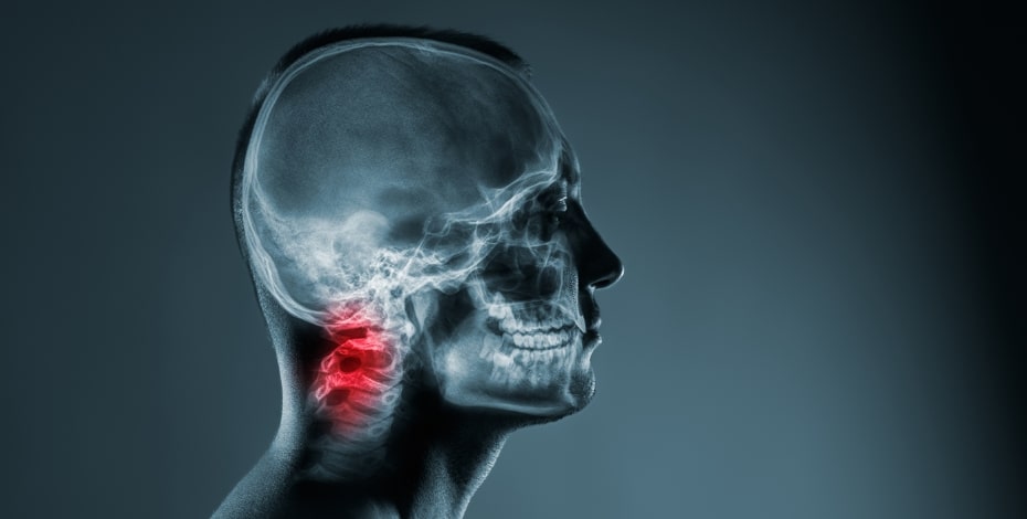
Cervical myelopathy is spinal cord dysfunction due to compression caused by narrowing of the spinal canal (McCartney et al 2018).
The pathophysiology of cervical cord compression (myelopathy) involves static and dynamic factors.
Static factors (eg, spondylosis, ligament hypertrophy and calcification) reduce the cross-sectional area of the spinal canal (Badhiwala et al 2020).
This effect is exacerbated by dynamic factors during pathological movement (eg, degenerative subluxation) and physiological movement (Badhiwala et al 2020).
The combination of these pathologies leads to chronic spinal compression, which can cause progressive neurological impairment and functional decline (Kalsi-Ryan et al 2012).
Furthermore, owing to the ageing population, rates of cervical myelopathy are increasing, along with resultant burden of disease (Badhiwala et al 2020).
Rapid diagnosis and treatment are imperative to avoid permanent disability (Badhiwala et al 2020).
Degenerative forms of cervical myelopathy are the most common cause of spinal cord impairment in adults worldwide (Kalsi-Ryan et al 2012).
Despite its prevalence, delay in diagnosis is common due to subtle early clinical features (Davies et al 2018), highly variable disease course and broad differential diagnoses (Fehlings & Mizuno 2018).
Diagnosis requires agreement between clinical and imaging findings (Badhiwala et al 2020).
Clinical manifestations may include neck pain or stiffness; pain, weakness or numbness in the upper or lower limbs; loss of manual dexterity; gait imbalance; and sphincter disturbance.
Magnetic resonance imaging (MRI) can distinguish the structural anatomical source and severity of spinal cord compression (Badhiwala et al 2020).
Detecting cervical myelopathy early can be challenging.
Differential diagnosis is further complicated in the presence of existing upper motor neuron (UMN) signs in stroke, a condition also more common in those aged 50 years and older.
The prevalence of upper limb impairment following stroke is approximately 50–80 per cent in the acute phase and 40–50 per cent in the chronic phase (Kwakkel et al 2003).
Commonly observed upper limb impairments after stroke are paresis, abnormal muscle tone and decreased somatosensation and coordination.
These deficits may also be found in cervical myelopathy, highlighting overlap with other neurological conditions and challenging timely and accurate diagnosis.
A consideration of the natural history of disease progression can assist with the differential diagnosis of cervical myelopathy concurrent with complex, co-existing UMN signs.
Longitudinal studies have repeatedly demonstrated the time dependency of neurological recovery after stroke (van der Vliet et al 2020).
In contrast, the natural history of degenerative cervical myelopathy generally consists of stepwise neurological decline (Fehlings & Mizuno 2018).
Some patients remain static in symptoms while others develop progressive disability and require surgery (Ferriter et al 2014).
Surgical decompression halts the progression of deficits and improves long-term neurological function, disability and quality of life (Badhiwala et al 2020).
Severe permanent disability is therefore preventable by early detection and intervention, before irreversible spinal cord injury has occurred (Badhiwala et al 2020).
It is the responsibility of primary healthcare professionals (eg, physiotherapists and GPs) to recognise the signs and symptoms of cervical myelopathy, differentially diagnose it from other neurological syndromes and initiate appropriate clinical care (Badhiwala et al 2020).
Case summary
The COVID-19 pandemic has significantly impacted healthcare.
The restrictions imposed to contain the spread of infection has limited access to health services, including rehabilitation.
Patients who have not received care due to the closure of outpatient services suffered collateral damage (Boldrini et al 2020).
This complex case demonstrates the high index of practitioner suspicion necessary when confronted with potential symptoms of cervical myelopathy on a background of chronic stroke.
A 61-year-old man with left spastic hemiplegia secondary to a stroke three years prior had recently been injected with botulinum neurotoxin type-A (BoNT-A) to address increasing spasticity (May 2020, Table 1).
He was attending twice-weekly physiotherapy complemented with a daily home exercise program targeted towards improving mobility and upper limb function.
He had made significant gains after the stroke to return to part-time work, driving and playing golf (Table 1).
This was his first experience with BoNT-A and he was highly motivated to improve his golf swing and walking endurance.
Eight weeks following the BoNT-A injection, his goals included being able to maintain his left hand grip on the golf club in mid-to-late swing, independent and unaided outdoor walking for up to four kilometres and an increased six minute walk test distance.
In early August 2020, COVID-19 restrictions reduced access to hospital-based outpatient services across Victoria, interrupting centre-based therapy.
Table 2 details the patient’s physical examination results (July 2020) prior to outpatient service closure (August 2020).
Telehealth services were offered but were declined.
The capability to remain motivated to perform regular independent home exercises had been demonstrated.
Therefore, the patient and therapist were hopeful functional gains could be maintained during lockdown.
Weekly welfare checks via telephone were conducted.
One week following cessation of centre-based therapy, the patient noticed a deterioration in upper limb function and increasing tightness in the forearm, reported via phone check-in.
BoNT-A interferes with neuromuscular synaptic transmission for 12 to 16 weeks following injection (Olver et al 2010).
It was deduced that increased tightness and reduced left arm function was most likely due to BoNT-A wearing off.
An urgent appointment for reinjection was initiated.
Physical examination (August 2020, mid-COVID-19 lockdown)
The spasticity clinic was allowed to review priority clients at risk of severe deterioration during lockdown (18 August 2020, Table 3).
The considerable increase in upper and lower limb spasticity and associated decline in upper limb functional activities and walking speed noted at reassessment drove the decision to further inject the left upper and lower limb musculature (Table 3) with BoNT-A.
Treatment goals were to increase self-selected walking speed and reduce the difficulty of opening jars and in performing reaching tasks.
The daily home program continued and weekly home-based exercise physiology sessions were initiated and included strength training (lower limbs), stretching, gait retraining, repetitive task practice and facilitated reach to grasp.
An eight-week telehealth review appointment was scheduled.
Outcome measures (October 2020, post-COVID-19 lockdown)
The International Classification of Functioning, Disability and Health (ICF) framework (WHO 2001) was applied to assessment and management.
Outcome measures relevant to stroke and functional decline
Patient complaints regarding further functional decline directed the physical examination and outcome measure selection.
Grip dynamometry and manual muscle testing (Kendall et al 1993) were performed to assess muscle power.
Muscle tone was assessed using the Modified Tardieu Scale (Boyd & Graham 1999) as it can differentiate spasticity from soft tissue hypertonia (Williams et al 2015).
Observation of upper and lower limb functional tasks was completed, including hand manipulation tasks, reach to grasp and button performance.
The nine-hole peg test measured dexterity and fine motor function (Wade 1989).
Evidence supports the use of the 10 metre walk test to measure gait speed and the six minute walk test to assess walking distance (Moore et al 2018); both were applied.
Outcome measures and diagnostic tests in suspicion of an alternative disease process
Given the unexpected decline in October 2020 and new symptoms, additional outcome measures were selected to aid differential diagnosis.
Subjective questioning relating to numbness, tingling, pain, weakness, loss of dexterity and difficulties with activities of daily living were explored.
Rapid alternating movements of hands and arms evaluated dysdiadochokinesia.
Eyes open and closed finger to nose coordination testing was performed to assess sensory ataxia.
A Romberg’s test assessed lower limb proprioception, indicating posterior column dysfunction (Emery 2001).
Cervical spine range of movement was measured.
Neural provocation tests of the upper limb were completed, along with a carpal tunnel compression test (Durkan 1991).
Physical examination (October–December 2020 post-COVID-19 lockdown)
Subjective examination
Initial reassessment (15 October 2020, Table 2 and Table 3) revealed that the patient had not participated in face-to-face physiotherapy or occupational therapy for nine weeks.
He had been seen by the exercise physiologist at home once a week.
He reported left upper limb functional deterioration with significant difficulty modulating grasp strength and controlling his wrist position.
He had lost the ability to lift the teapot lid, scoop muesli and apply deodorant using the left hand.
Due to COVID-19 restrictions, the patient was unable to play his regular twice-a-week golf and his walking distance had reduced.
He had also recently lost three kilograms and was looking very thin.
The patient was feeling discouraged regarding the lack of progress in the left arm since the BoNT-A injection in August 2020.
Once-a-week physiotherapy for stroke management was re-initiated (October–December 2020) following the reassessment.
During this period of rehabilitation, an alternative disease process was considered (Table 2 and Table 3).
Key subjective reports during the patient’s weekly physiotherapy (October–December 2020, Table 3) included ongoing vague sensory changes, fluctuating left arm tightness, inability to relax the arm and hand when lying flat and ongoing difficulty gripping objects.
Of note were 3–4 episodes of left upper limb dystonia and numbness that occurred when straining or stressing the arm, with symptoms improving on lying down within one hour.
One instance had resulted in a presentation to the emergency department (23 November 2020) where an MRI of the brain ruled out arteriovenous malformation progression or stroke.
Impairment and activity measures
The key impairment test results (Table 2 and Table 3) that created a high level of suspicion included new dystonic posturing of the left thumb adductors, reduced coordination testing with the eyes closed (finger nose (FN) eyes open (EO) 6/10sec; eyes closed (EC) 3/10sec), inability to maintain wrist extension during finger extension and deterioration of rapid alternating movements with visible tremor.
The nine-hole peg test could no longer be performed.
There was increased intrinsic muscle wasting, along with reduced grip strength (22.35kg compared with 25.76kg).
The patient could no longer perform a single leg bridge on the left.
There was mild trunk ataxia on double leg bridging.
The Romberg’s test was newly positive and he now required a single point stick outdoors.
Physical examination continued (October–December 2020 post-COVID-19 lockdown)
Differential diagnosis—clinical reasoning process
Figure 1 outlines the differential diagnosis process.
The treating physiotherapist advocated an MRI of the brain and spine via the rehabilitation consultant (19 November 2021).
Knowledge of the natural history of chronic stroke (predictable pattern of time-dependent recovery) greatly assisted the differential diagnosis process.
Cervical myelopathy often progresses with acute episodic exacerbations causing the subsequent decline in motor function (Vigna & Tortolani 2004).

Elizabeth Moore's case study investigates cervical myelopathy on a background of chronic stroke.
Hence, the presence of new UMN signs superimposed upon previous UMN signs with significant loss of upper limb function were indicative of a different underlying disease process than physical deconditioning alone following lockdown.
Although, the nine-week period of reduced activity contributed to reduced strength, decreased muscle size and muscle atrophy, new sensory and proprioceptive changes and fluctuating dystonia suggested an alternative diagnosis.
Subtle lower motor neuron (LMN) signs raised red flags for potential peripheral nervous diseases.
Amyotrophic lateral sclerosis (ALS) and cervical myelopathy typically present in older adults.
The neurological examination in both disorders often demonstrates mixed UMN and LMN deficits.
With cervical myelopathy, LMN deficits and fasciculations are isolated to the affected cervical myotomes, but in ALS they also often appear in the legs and cranial muscles (eg, tongue; Vigna & Tortolani 2004).
Sensory deficits are not expected in ALS, thus ALS was eliminated.
A brain or spinal tumour was considered in light of the new neurological symptoms and recent three-kilogram weight loss.
An MRI of the brain (23 November 2020) ruled out a brain tumour and stroke progression as potential causes.
Injections with botulinum toxin are generally well tolerated and side-effects are few (Nigam & Nigam 2010).
Its effect diminishes with increasing distance from the injection site, but spread to nearby muscles is possible (Nigam & Nigam 2010).
The reduction in strength observed in the wrist extensors and grip muscles and wasting of the intrinsic muscle could not have been caused by BoNT-A spread as these muscles do not border the injected muscles.
Carpal tunnel was eliminated as a potential diagnosis following a carpal tunnel compression test.
Proximal nerve entrapment around the shoulder and other shoulder impairment may have contributed to the reduced arm strength and sensory changes.
A shoulder MRI (10 December 2020) confirmed a chronic superior lesion anterior to posterior tear and although a likely contributor to the reduced shoulder strength, it would not explain the more distal neurological symptoms.
A spinal tumour was eliminated following an MRI of the spine (10 December 2020).
Confirmed diagnosis
MRI findings correlated with the clinical assessment and confirmed a diagnosis of multilevel, C3/C4, C4/C5 and C5/C6, mild-to-moderate cervical cord compression with focal chronic myelomalacia, secondary to focal disc protrusions and severe canal stenosis.
Management plan and outcome
Posterior cervical laminectomy and fusion of C3–C7 was performed, aiming to halt progression.
However, due to the dynamic component to the symptoms, there was hope of improvement.
Immediately after the operation the patient noticed improvement in both upper and lower limbs.
He was discharged from inpatient rehabilitation 15 days post-surgery and recommenced outpatient therapy (February 2021).
Six weeks following the operation his grip strength had increased (27.7kg compared with 22.35kg) and he was able to perform the nine-hole peg test (9/9 4min 27sec compared with 3/9 unfinished test).
Upper limb sensory ataxia persisted (FN EO 4/10, EC 2/10).
However, he no longer required a single point stick outdoors and could walk up to three kilometres unaided (Table 3).
Discussion
Stroke is considered a non-progressive acquired neurological disorder.
Recovery from stroke follows a time-dependent predictable pattern (Kwakkel et al 2004) with a slow but steady upwards trajectory.
However, with the majority of strokes occurring in people 65 years or older, existing neurological symptoms may change with potential additional neurological aetiologies, such as Parkinson's disease, ALS or Alzheimer's disease.
These people are at risk of remaining undiagnosed or undertreated.
No studies have investigated the incidence of new onset neurological issues in the chronic stroke population.
A recent review found adults with cerebral palsy have an increased risk of new neurological conditions, such as stroke and myelopathy, which led to the development of guidelines for longitudinal neurological screening (Smith et al 2021).
The diagnostic process in this case highlights how clinical signs and symptoms of cervical myelopathy may be obscure, particularly when cervical myelopathy can masquerade as another disease process (Vigna & Tortolani 2004).
This study demonstrates the need for ongoing neurological surveillance in chronic stroke populations, such as recommended in cerebral palsy, particularly as new motor impairments can be difficult to distinguish from longstanding impairments without rigorous neurological examination tracking (Smith et al 2021).
Cervical myelopathy can be elusive to diagnose (Rumi & Yoon 2004) and this is in the absence of competing existing UMN signs.
Objective physical indicators of cervical myelopathy include UMN signs (eg, hyperreflexia, clonus, a positive Babinski sign and spasticity).
These findings mimic symptoms the patient, in this case, was already experiencing from a previous stroke.
When working with chronic stroke patients, clinicians may observe symptoms through a 'stroke lens' and not be attuned to a broader range of neurological assessments.
The finger escape sign (Rumi & Yoon 2004) and vibration testing would have added value to the physical assessment in this case.
Clinicians need to consider an expanded set of assessment tools when a patient doesn't 'fit the pattern' of a prior diagnosis.
People with chronic stroke are prone to functional decline with a reduction in physical activity (Billinger et al 2014).
The COVID-19 pandemic has had a negative impact in the short term, with functional deterioration in persons with chronic diseases (Boldrini et al 2020).
Hence, it was reasonable to attribute the initial functional decline in this case to a lack of therapy.
Additionally, reduced upper limb strength following BoNT-A injection was possible.
However, all diagnostic explanations must be considered when presented with new and emerging symptoms.
This case highlights the important role of physiotherapists in applying clinical reasoning skills and advocating for further investigations in complex patients.
Early detection of cervical myelopathy is accomplished clinically and confirmed radiographically.
Features can be mild and difficult to elicit in the initial stages of disease.
As a result, average delays of 2.2 years (range 1.7 months to 8.9 years) from initiation of symptoms to diagnosis have been reported (Behrbalk et al 2013).
In the presence of coexisting UMN signs from a stroke, this time frame would undoubtedly increase.
The time to diagnose cervical myelopathy in this case, from presentation of initial symptoms, was less than two months.
This case endorses the value of an expeditious diagnosis and treatment of cervical myelopathy to avoid permanent disability (Badhiwala et al 2020).
The main limitation of this study is that findings are applicable to the case in question, and thus cannot be generalised more widely.
There is also the possibility of a risk of bias from uncontrolled observations; therefore results should be interpreted with caution.
This study, however, provides useful information regarding clinician diagnostic reasoning in a complex presentation of new UMN signs on a background of chronic stroke.
Patient consent has been provided for this case to be published.
>> Elizabeth Moore, MACP, is a registrar undertaking Fellowship of the Australian College of Physiotherapists by Clinical Specialisation in the neurology discipline. Elizabeth is a senior physiotherapist and Neurology and Spasticity Clinic Team Leader at Epworth HealthCare.
- References
McCartney, S., Baskerville, R., Blagg, S., & McCartney, D. (2018). Cervical radiculopathy and cervical myelopathy: diagnosis and management in primary care. British Journal of General Practice, 68(666), 44-46.
Badhiwala, J. H., Ahuja, C. S., Akbar, M. A., Witiw, C. D., Nassiri, F., Furlan, J. C., . . . Fehlings, M. G. (2020). Degenerative cervical myelopathy—update and future directions. Nature Reviews Neurology, 16(2), 108-124.
Kalsi-Ryan, S., Karadimas, S. K., & Fehlings, M. G. (2012). Cervical Spondylotic Myelopathy: The Clinical Phenomenon and the Current Pathobiology of an Increasingly Prevalent and Devastating Disorder. The Neuroscientist, 19(4), 409-421. doi:10.1177/1073858412467377
Davies, B. M., Mowforth, O. D., Smith, E. K., & Kotter, M. R. (2018). Degenerative cervical myelopathy. BMJ, 360, k186. doi:10.1136/bmj.k186
Fehlings, M. G., & Mizuno, J. (2018). Cervical Myelopathy, An Issue of Neurosurgery Clinics of North America, E-Book.
Kwakkel, G., Kollen, B. J., van der Grond, J., & Prevo, A. J. (2003). Probability of regaining dexterity in the flaccid upper limb: impact of severity of paresis and time since onset in acute stroke. Stroke, 34(9), 2181-2186.
van der Vliet, R., Selles, R. W., Andrinopoulou, E. R., Nijland, R., Ribbers, G. M., Frens, M. A., . . . Kwakkel, G. (2020). Predicting upper limb motor impairment recovery after stroke: a mixture model. Annals of neurology, 87(3), 383-393.
Ferriter, P. J., Mandel, S., Degregoris, G., Kamara, E., & AYDIN, S. M. (2014). Cervical myelopathy. Practical Neurology, 43-46.
Boldrini, P., Garcea, M., Brichetto, G., Reale, N., Tonolo, S., Falabella, V., . . . Kiekens, C. (2020). Living with a disability during the pandemic." Instant paper from the field" on rehabilitation answers to the COVID-19 emergency. European journal of physical and rehabilitation medicine.
Olver, J., Esquenazi, A., Fung, V. S. C., Singer, B. J., & Ward, A. B. (2010). Botulinum toxin assessment, intervention and aftercare for lower limb disorders of movement and muscle tone in adults: International consensus statement. European Journal of Neurology, 17(SUPPL. 2), 57-73.
santé, O. m. d. l., Organization, W. H., & Staff, W. H. O. (2001). International classification of functioning, disability and health: ICF: World Health Organization.
Kendall, F. P., McCreary, E. K., Provance, P. G., Rodgers, M., & Romani, W. A. (1993). Muscles, testing and function: with posture and pain (Vol. 103): Williams & Wilkins Baltimore, MD.
Boyd, R. N., & Graham, H. K. (1999). Objective measurement of clinical findings in the use of botulinum toxin type A for the management of children with cerebral palsy. European Journal of Neurology, 6, s23-s35.
Williams, G., Banky, M., & Olver, J. (2015). Severity and distribution of spasticity does not limit mobility or influence compensatory strategies following traumatic brain injury. Brain injury, 29(10), 1232-1238.
Wade, D. T. (1989). Measuring arm impairment and disability after stroke. International disability studies, 11(2), 89-92.
Moore, J. L., Potter, K., Blankshain, K., Kaplan, S. L., O'Dwyer, L. C., & Sullivan, J. E. (2018). A core set of outcome measures for adults with neurologic conditions undergoing rehabilitation: a clinical practice guideline. Journal of Neurologic Physical Therapy, 42(3), 174.
Emery, S. E. (2001). Cervical spondylotic myelopathy: diagnosis and treatment. JAAOS-Journal of the American Academy of Orthopaedic Surgeons, 9(6), 376-388.
Durkan, J. A. (1991). A new diagnostic test for carpal tunnel syndrome. J Bone Joint Surg Am, 73(4), 535-538
Vigna, F. E., & Tortolani, P. J. (2004). Cervical myelopathy: Differential diagnosis. Paper presented at the Seminars in Spine Surgery.
Nigam, P. K., & Nigam, A. (2010). Botulinum toxin. Indian journal of dermatology, 55(1), 8-14. doi:10.4103/0019-5154.60343
Smith, S. E., Gannotti, M., Hurvitz, E. A., Jensen, F. E., Krach, L. E., Kruer, M. C., . . . Aravamuthan, B. R. (2021). Adults with cerebral palsy require ongoing neurologic care: a systematic review. Annals of neurology, 89(5), 860-871.
Rumi, M. N., & Yoon, S. T. (2004). Cervical myelopathy history and physical examination. Paper presented at the Seminars in Spine Surgery.
Behrbalk, E., Salame, K., Regev, G. J., Keynan, O., Boszczyk, B., & Lidar, Z. (2013). Delayed diagnosis of cervical spondylotic myelopathy by primary care physicians. Neurosurgical focus, 35(1), E1.
© Copyright 2025 by Australian Physiotherapy Association. All rights reserved.


